Spinal Imaging
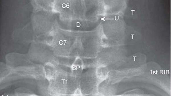
It may be suggested after clinical assessment that you require imaging of your spine. Commonly used investigations are standing x-rays to look at overall balance. Occasionally dynamic x-rays can be used to show abnormal movement/instability.Further investigations may include an MRI scan (Magnetic Resonance Imaging) which details the discs and nerves more closely. A CT (Computerised Tomography) offers a more detailed look at bony architecture and can be used after fusion surgery to assess fusion. It does involve x-rays unlike an MRI scan. Bone scans or SPECT scans can be used to look more closely at areas of pain and when there are suspicions of more serious pathology.
DEXA scans can be used to assess bone density in patients with suspicions of osteoporosis. Nerve conduction tests or electromyography can be used to assess whether there is a nerve problem in the upper or lower limb, and it can also assess muscle function. This test is done by a neurologist. Occasionally discography can be used to try and identify a pain source from a disc and this is a radiological test where a needle is placed using x-ray guidance into the disc and x-ray dye is inserted into the disc to increase the pressure within the disc to see if that reproduces a localised concord back pain.
Pease see more information on Spinal Imaging